
A technical deep-dive into the cellular mechanisms behind photobiomodulation (PBM) technology — the field underpinning red and near-infrared light therapy devices.
Chapter 1: A Brief History — From Accidental Discovery to a Field of 6,000+ Studies
The story of photobiomodulation begins not with a planned experiment, but with a mistake.
In 1967, Hungarian physician and surgeon Endre Mester was working at Semmelweis University in Budapest. Inspired by earlier research showing that high-powered lasers could destroy tumours in rats, he set out to test whether lasers might cause cancer in mice. He shaved the backs of two groups of mice and applied a low-powered ruby laser (694nm, 1mW) to one group. Cancer did not develop in either group. What Mester didn't expect was what happened next: the hair on the laser-treated mice grew back significantly faster than on the untreated controls. He had stumbled onto what he called "laser biostimulation" — the stimulatory effect of low-level light on biological tissue, without thermal damage.[1]
Mester went on to publish extensively on wound healing and tissue regeneration using low-level laser light. His work seeded an entire field. Over the following decades — largely through the separate but equally foundational research of Russian biophysicist Tiina Karu — the molecular mechanisms began to be identified. Karu's work in the 1980s and 1990s was instrumental in pinpointing cytochrome c oxidase as the primary cellular photoreceptor, shifting the field from empirical observation toward mechanistic understanding.[2]
The 1990s brought two significant technological shifts. First, efficient red and near-infrared LEDs became commercially viable — enabling large-area coverage that was impractical with lasers. Second, NASA-funded research by Harry Whelan and colleagues began exploring LED-based photobiomodulation for wound healing in microgravity environments, lending institutional credibility and generating data that fed directly into clinical protocols still in use today.
By the 2000s, the field had matured enough to shed its early nomenclature. What had been called variously "laser biostimulation," "low-level laser therapy (LLLT)," "cold laser therapy," and "soft laser therapy" was standardised under the term photobiomodulation (PBM) — a name that reflects the mechanism (modulation of biological processes by light) rather than the hardware used to deliver it.
Today, PBM research spans more than 6,000 peer-reviewed studies indexed in PubMed, examining everything from cellular energy metabolism to applications in dermatology, musculoskeletal research, and neuroscience. The term "LLLT" remains in common use and refers to the same field.
Chapter 2: What Is Photobiomodulation? Defining the Technology
Photobiomodulation is the use of light at specific, non-ionising wavelengths — typically in the red (620–700nm) and near-infrared (700–1100nm) portions of the electromagnetic spectrum — to interact with chromophores inside cells and modulate cellular activity.[3]
A few definitional boundaries are worth setting clearly:
It is not heat therapy. PBM operates at irradiance levels that do not meaningfully raise tissue temperature. This distinguishes it from infrared saunas, heat lamps, or far-infrared devices, which work through thermal mechanisms. The cellular effects of PBM are photochemical — driven by photon absorption — not thermal.
It is not photodynamic therapy. Photodynamic therapy uses light to activate an externally applied photosensitising agent. PBM uses no external agent; it interacts with naturally occurring chromophores already present in cells.
It is not UV or ionising radiation. The wavelengths used in PBM sit well below the ultraviolet range. They do not damage DNA and are not associated with the carcinogenic risks of UV exposure.
It does not operate through placebo-like non-specific effects. The cellular mechanisms are measurable, reproducible in vitro (in isolated cells and tissues), and have been identified at the molecular level. The primary photoreceptor — cytochrome c oxidase — has been characterised in detail across multiple independent research programmes.
What PBM does is deliver photons at wavelengths that are absorbed by specific molecular structures inside cells, triggering a downstream cascade of biochemical events. The nature of those events — and why the wavelength and dose must be within specific parameters for the cascade to occur — is the subject of the chapters that follow.
Chapter 3: The Primary Mechanism — Cytochrome c Oxidase and Mitochondrial Energy Production
The central mechanism of PBM is well-established in the peer-reviewed literature: red and near-infrared photons are absorbed by cytochrome c oxidase (CCO), a key enzyme in the mitochondrial electron transport chain, stimulating increased production of adenosine triphosphate (ATP) — the molecule cells use as their primary energy currency.
To understand why this matters, it helps to understand what cytochrome c oxidase actually does.
The Electron Transport Chain and ATP Synthesis
Mitochondria generate ATP through a process called oxidative phosphorylation. The electron transport chain (ETC) is a series of protein complexes embedded in the inner mitochondrial membrane. Electrons are passed along these complexes, and the energy released is used to pump protons (H⁺ ions) across the membrane, creating a gradient. This gradient drives ATP synthase — the molecular turbine that converts the proton gradient into ATP.
Cytochrome c oxidase is Complex IV of the ETC — the final enzyme in the chain. It accepts electrons from cytochrome c and transfers them to oxygen, producing water. This step is the rate-limiting reaction of the entire process: when CCO activity is high, the ETC runs efficiently and ATP production is elevated. When CCO activity is suppressed — for example, by nitric oxide occupying its active site — the chain slows, and ATP output falls.
How Light Interacts with CCO
CCO contains metallic chromophores — specifically copper centres (CuA and CuB) and haem groups (haem a and haem a3) — that absorb photons in the red and near-infrared range. When a photon is absorbed by one of these chromophores, it changes the enzyme's redox state, enhancing its catalytic activity.
Research published in the Journal of the American Academy of Dermatology (2024) describes the process precisely: red and near-infrared light stimulates CCO, leading to upregulation of the electron transport chain, which results in increased mitochondrial ATP production.[4]
A pivotal aspect of this mechanism involves nitric oxide (NO). Under cellular stress, NO — a freely diffusible signalling molecule — can bind to CCO and competitively inhibit it, displacing oxygen and reducing ATP output. PBM-induced photon absorption is understood to photodissociate this bound NO, freeing CCO to resume normal oxygen binding and resuming full ATP synthesis.[5] The released NO then enters the bloodstream as a free signalling molecule, where it has its own downstream effects (discussed in Chapter 4).
The ATP Cascade
Research from Cardoso et al. (2022), published in Frontiers in Neuroscience, confirms that CCO is the major intracellular acceptor of photons in the red to near-infrared wavelengths used for PBM, and that this absorption promotes changes in the redox state of enzymes in the mitochondrial inner membrane, improving energy metabolism through increased ATP synthesis.[6]
A comprehensive review (ScienceDirect, 2020) frames it clearly: PBM targets cytochrome c oxidase, leading to increased synthesis of adenosine triphosphate, induction of growth factor secretion, activation of signalling pathways, and promotion of gene expression.[7]
When cells have more available ATP, they perform their functions more efficiently. This is the foundational principle from which the broader downstream effects of PBM follow.
Chapter 4: The Secondary Cascade — What Happens After ATP Rises
Increased ATP production is the primary event in PBM, but it initiates a broader intracellular signalling cascade. Understanding these secondary effects is important for appreciating why PBM research spans such a wide range of applications.
Reactive Oxygen Species (ROS) as Signalling Molecules
PBM at appropriate doses generates a transient, mild increase in reactive oxygen species (ROS) within mitochondria. This sounds counterintuitive — ROS are often discussed in the context of oxidative stress. But at low-to-moderate concentrations, ROS are well-established redox signalling molecules that activate downstream pathways involved in cellular repair, proliferation, and adaptation.
It is only at high concentrations — well above the doses produced by PBM — that ROS become cytotoxic. The dose-dependency of this response is a key reason why irradiance parameters matter in PBM research, and why the biphasic dose-response relationship (discussed in Chapter 6) is so important.
Nitric Oxide Release and Microcirculation
As described in Chapter 3, PBM-induced photodissociation of NO from CCO releases free nitric oxide into the cell and surrounding tissue. NO is a potent vasodilator — a signalling molecule that relaxes the smooth muscle of blood vessel walls, causing them to expand.
A 2022 review in Nitric Oxide (Kashiwagi et al.) confirms that photon absorption at wavelengths between 600 and 1,100nm enhances enzymatic activity, increasing ATP production and transiently releasing NO, which contributes to vasodilation and improved microcirculation.[8]
A 2024 clinical study published in Photodiagnosis and Photodynamic Therapy measured NO release from intact human skin following exposure to 660nm and 850nm wavelengths in 18 healthy participants, providing direct in-vivo evidence of this release mechanism.[9]
Transcription Factor Activation
The changes in cellular redox state triggered by PBM activate a range of transcription factors — proteins that regulate gene expression. Key among these is NF-κB (nuclear factor kappa-light-chain-enhancer of activated B cells), which plays a central role in modulating inflammatory responses, and AP-1, which is involved in cell proliferation and differentiation.
A 2024 review in PMC on the immunomodulatory effects of PBM confirms that the boost in cellular energy availability following CCO stimulation has profound effects on various cellular processes, including immune responses, through these downstream signalling mechanisms.[10]
Calcium Ion Flux
PBM has also been shown to influence intracellular calcium (Ca²⁺) signalling. Calcium ions act as secondary messengers in a wide range of cellular processes. The changes in mitochondrial membrane potential triggered by PBM affect Ca²⁺ flux, contributing to the activation of downstream repair and proliferation pathways.
This multi-pathway cascade — ATP, ROS signalling, NO release, transcription factor activation, calcium flux — explains why PBM research explores such a broad range of physiological applications from a single cellular entry point.
Chapter 5: Wavelength Science — Why 660nm and 850nm Specifically?
Not all red or infrared light triggers PBM responses. The mechanism is wavelength-dependent: photons must be at frequencies that correspond to the absorption peaks of cellular chromophores, primarily CCO. The research literature has converged on two wavelength ranges as particularly well-characterised: visible red around 660nm, and near-infrared around 850nm.
The Optical Window
Before examining the two wavelengths individually, it helps to understand the concept of the "optical window" — the range of wavelengths (approximately 600–900nm) in which light has the best combination of tissue penetration and cellular absorption.
- Below ~600nm (visible green, blue, UV): Strongly absorbed by haemoglobin and melanin. Very limited tissue penetration.
- Above ~1,000nm: Increasingly absorbed by water in tissue. Penetration falls off significantly.
- 600–900nm: The "optical window" — light penetrates tissue to useful depths while still being absorbed by chromophores in mitochondria.
Both 660nm and 850nm sit comfortably within this window, but at different positions with different penetration profiles.
660nm: Visible Red
At 660nm, light sits in the visible red portion of the spectrum. It is absorbed strongly by CCO's haem a3 copper centre and corresponds closely to one of CCO's primary absorption peaks.
Research consistently shows that 660nm light penetrates to approximately 2–5mm into tissue, reaching the epidermis and dermis — the layers where fibroblasts (the cells responsible for collagen synthesis), keratinocytes, and their associated mitochondria are most concentrated.[11] Kolárová et al. (1999, Journal of Photochemistry and Photobiology B) measured actual light transmission through human tissue samples and found that 660nm light retained approximately 5–10% of surface intensity at 5mm depth in skin, dropping below 1% at 10mm — consistent with the dermis as its primary zone of influence.[12]
The 660nm wavelength has accumulated the largest volume of published clinical research of any single visible red wavelength, with trials across wound healing, skin fibroblast activity, and collagen production protocols repeatedly using this specific frequency.
850nm: Near-Infrared
At 850nm, light crosses out of the visible spectrum entirely. The human eye cannot detect 850nm light, though camera sensors can often register a faint glow from NIR LEDs.
The longer wavelength means reduced scattering and absorption in tissue, giving 850nm a substantially deeper penetration profile compared to 660nm. Research literature reports penetration depths ranging from 30mm to over 50mm depending on tissue composition, with the practical therapeutic range for musculoskeletal applications typically cited at 3–5cm from the skin surface.[13]
At 850nm, CCO absorption remains significant, but the deeper penetration shifts the primary zone of interaction toward muscle tissue, connective tissue, and joints rather than the superficial skin layers. This is why research examining PBM in the context of deep tissue applications overwhelmingly favours NIR wavelengths over visible red.
Why Use Both?
The complementary penetration profiles of 660nm and 850nm are why dual-wavelength devices have become the standard in serious PBM research and device design. The two wavelengths work in parallel:
- 660nm drives interaction at the dermis and superficial tissue level, where the skin's cellular architecture and collagen-producing fibroblasts reside.
- 850nm reaches deeper tissue structures that 660nm cannot reliably access.
The evidence supports the complementary nature of the two wavelengths. Research using both simultaneously has generally produced results consistent with additive or synergistic effects, rather than one wavelength simply replicating the other.
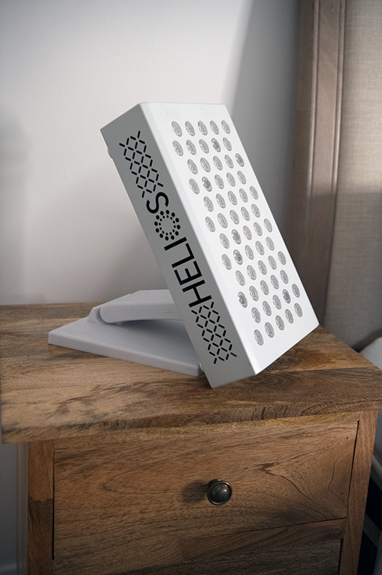
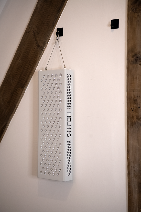
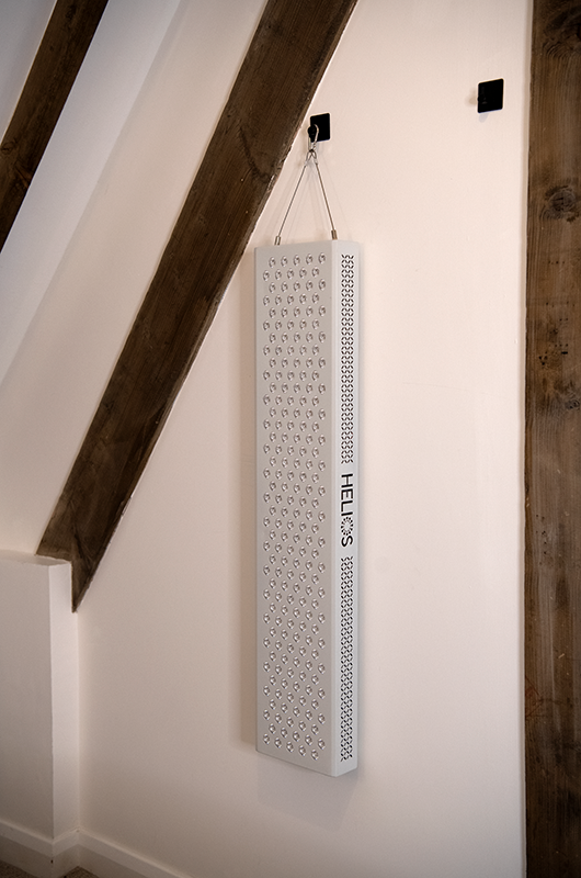
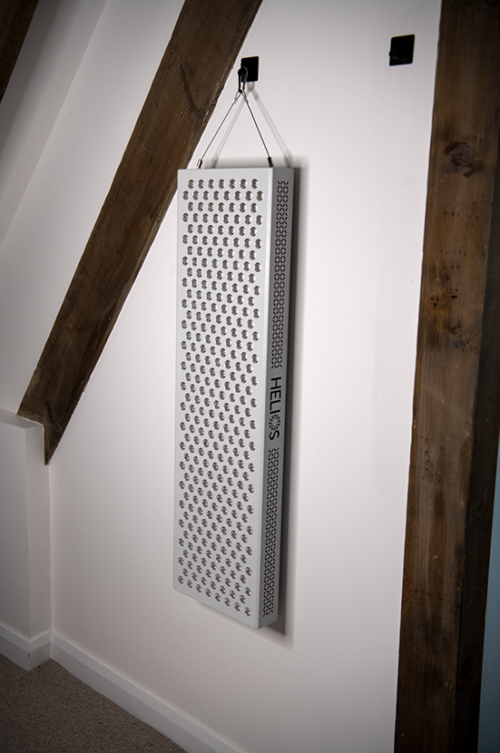
HELIOS 2 Series devices emit both 660nm and 850nm simultaneously by default, covering both penetration depths in a single session.
Chapter 6: Irradiance, Fluence, and the Dose-Response Relationship
Understanding PBM requires understanding the physics of light dosimetry. Two parameters are foundational:
Irradiance (measured in mW/cm²): the power of light delivered per unit area. This is what device specifications express — for example, ">135 mW/cm² at 6 inches" for the HELIOS 2 1500W panel. Higher irradiance means more photons delivered per second.
Fluence (measured in J/cm², also called energy density or dose): irradiance multiplied by time. This is the total energy delivered to a unit area over a session. A device delivering 100 mW/cm² for 10 minutes produces a fluence of 60 J/cm².
Both parameters matter, and neither alone determines whether PBM will be effective.
The Arndt-Schulz Curve: Why More Is Not Always Better
One of the most important — and frequently misunderstood — aspects of PBM is the biphasic dose-response relationship, formally described by the Arndt-Schulz law.
Research by Huang, Sharma, Carroll, and Hamblin (2009, updated 2011 in Dose-Response) established this relationship comprehensively: too little light produces insufficient cellular response; an optimal dose range produces maximum biological activity; too much light produces a plateau and, at excessive doses, inhibitory effects.[14]
A 2025 paper in ScienceDirect confirms: the biphasic dose-response (Arndt-Schulz law) is a fundamental characteristic of PBM, in which both insufficient and excessive energy densities reduce efficacy, with maximal biological effects achieved within an optimal dose range.[15]
This is fundamentally unlike pharmaceutical dosing, where the common assumption is monotonically increasing response. In PBM:
- Sub-threshold doses (typically below ~1–2 J/cm²): Insufficient photon density to drive measurable CCO upregulation.
- Optimal range (approximately 3–50 J/cm² depending on tissue depth and target): Maximum ATP induction, ROS signalling, and downstream cascade activation.
- Supra-threshold doses (above ~60–100 J/cm²): Diminishing returns; at extreme doses, potential inhibitory effects have been observed.
The optimal range varies by tissue depth and application — surface tissue targets require less fluence than deep tissue targets, since delivered photons must be sufficient at depth, not just at the surface.
What This Means for Device Use
The biphasic dose response has a practical implication: session duration is not infinitely scalable. The goal is to land within the therapeutic window, not simply maximise light exposure. For most surface applications, 10–20 minutes at 6–12 inches from a panel operating at therapeutic irradiance (>100 mW/cm²) falls comfortably within the researched range.
This is also why irradiance specifications matter when evaluating devices: a device with inadequate irradiance may require impractically long sessions to reach the minimum effective fluence, particularly for deeper tissue targets.
Chapter 7: LED vs. Laser — Does Coherence Matter?
A common question in PBM discussions is whether the coherent (phase-aligned) light produced by lasers is superior to the non-coherent light from LEDs. The short answer, based on the current evidence, is: for the biological mechanisms of PBM, coherence does not appear to be the determining factor.
The critical variable is wavelength and delivered irradiance. The photon must be at a frequency that CCO absorbs, and must be delivered at sufficient density to drive the cascade. Whether those photons arrive in phase with one another is biologically irrelevant once they are absorbed by the chromophore.
Multiple research groups have compared LED and laser delivery at matched wavelengths and fluences and found equivalent biological outcomes. The shift from laser-based research to LED-based devices has not required a re-examination of the underlying mechanisms — the mechanisms are the same.
The practical advantages of LEDs are significant:
- Large treatment area: A single LED panel can illuminate the entire torso simultaneously. An equivalent laser treatment would require manual scanning and would be impractical for home use.
- Safety: LEDs operate at power densities that do not carry the retinal risk of Class 3B or 4 lasers.
- Cost: LED panels deliver therapeutic irradiance at a fraction of the cost of equivalent laser systems.
- Heat: LEDs at therapeutic power levels do not heat tissue, keeping the interaction photochemical rather than thermal.
This is the technological evolution Mester's original work eventually led to: from single-point laser probes treating small areas in clinical settings, to large-array LED panels capable of full-body illumination at home.
Chapter 8: The Optical Window — Why Red and NIR Light Penetrate Tissue
Why do 660nm and 850nm light penetrate tissue at useful depths while other wavelengths do not? The answer lies in the interaction of different wavelengths with tissue components.
Human tissue is a complex optical medium containing several major light-absorbing molecules (chromophores):
- Melanin: Absorbs strongly below ~700nm, with absorption falling rapidly through the red range. This is why visible red light (660nm) penetrates less than NIR.
- Oxyhaemoglobin and deoxyhaemoglobin: Absorb heavily below ~600nm and above ~1,000nm. In the red and NIR window, haemoglobin absorption is relatively low.
- Water: Absorbs progressively more above ~1,000nm. At 1,300nm+, water becomes a dominant absorber and penetration drops sharply.
- Lipids: Relatively transparent in the 600–1,000nm range.
The 600–900nm optical window is the zone where all four major tissue chromophores have relatively low absorption, allowing light to penetrate to clinically useful depths. Within this window, shorter wavelengths (600–700nm) still encounter moderate melanin absorption; longer wavelengths (800–900nm) have the better penetration profile.
Scattering — the deflection of photons by tissue structures rather than their absorption — is also a factor. Both absorption and scattering decrease as wavelength increases through the optical window, which contributes to the superior depth penetration of 850nm over 660nm.
The net result is the depth profiles described in Chapter 5: 660nm reaching 2–5mm primarily in the dermis; 850nm penetrating 3–5cm into deeper tissue structures.
Chapter 9: Device Specifications That Actually Matter
Understanding the underlying science helps cut through marketing claims when evaluating PBM devices. Here are the specifications that are directly relevant to the photobiomodulation mechanisms described in this article.
Wavelength (nm)
The most fundamental specification. The wavelength must fall within the absorption spectrum of CCO and within the tissue optical window. 660nm and 850nm are the two most extensively researched specific wavelengths in the PBM literature. Devices advertising proprietary wavelengths in the 700–800nm range — where absorption by CCO drops off — should be scrutinised against the published research.
Irradiance at Distance (mW/cm²)
This is the power delivered per unit area at a specified distance from the device. It determines whether a session of reasonable duration will deliver fluence within the therapeutic window. An irradiance below ~50 mW/cm² at typical treatment distances (6–18 inches) requires impractically long sessions; >100 mW/cm² at 6 inches is typically cited in serious device specifications.
The HELIOS 2 1500W panel delivers >135 mW/cm² at 6 inches, >107 mW/cm² at 12 inches, and >85 mW/cm² at 18 inches — all within the range where meaningful fluence accumulates in 10–20 minutes.
Rated Power and Power Consumption
These are distinct figures. "Rated power" in LED panels refers to the total LED wattage — the sum of all individual LED ratings. "Power consumption" is actual electrical draw. For the HELIOS 2 range:
| Device | Rated Power | Power Consumption | LED Count |
|---|---|---|---|
| 300W | 300W | 91W | 60 × 5W LEDs |
| 600W | 600W | 215W | 120 × 5W LEDs |
| 1000W | 1000W | 302W | 200 × 5W LEDs |
| 1500W | 1500W | 375W | 300 × 5W LEDs |
The gap between rated and consumed power is standard in the industry and reflects that LEDs are never driven at their maximum rated wattage; running at 20–30% of rated wattage extends LED lifespan significantly while still delivering therapeutic irradiance.
EMF Emissions
HELIOS 2 devices are tested at 0.0 µT at 6 inches — below detectable electromagnetic field levels at treatment distance. This is relevant for users who are attentive to electromagnetic exposure.
Light Angle (Degrees)
The 60-degree light angle on HELIOS 2 panels is an optical design decision affecting how light is distributed across a treatment area. Narrower angles concentrate output in smaller areas with less edge scatter; wider angles increase coverage area at the cost of intensity at centre.
Chapter 10: The Current State of the Research Literature
PBM sits in an interesting position in the research landscape. The cellular mechanisms — CCO stimulation, ATP production, ROS signalling, NO release — are well-characterised and reproducible at the molecular level. The volume of published research exceeds 6,000 indexed studies. Several systematic reviews and meta-analyses exist across specific application areas.
At the same time, the field faces genuine methodological challenges. Many early studies used small sample sizes, varied widely in delivery parameters (making cross-study comparisons difficult), and lacked appropriate controls. The heterogeneity of parameters — wavelength, irradiance, fluence, treatment area, application protocol — has historically made it difficult to derive consistent clinical conclusions.
The field has responded to these challenges through better standardisation. The work of researchers such as Michael Hamblin (Harvard Medical School), Tiina Karu, Juanita Anders, and the broader NAALT (North American Association for Light Therapy) community has pushed for tighter dosimetric reporting, consistent terminology, and more rigorous trial design.
The 2024 review in the Journal of the American Academy of Dermatology (Mineroff et al.) represents the kind of peer-reviewed synthesis now appearing in mainstream clinical literature — applying standardised assessment frameworks to the PBM evidence base.[4]
For a technology once dismissed as fringe, PBM's trajectory over the past decade — with publication rates accelerating, systematic reviews accumulating, and dermatology's main professional journal publishing CME-accredited coverage — represents a meaningful shift in scientific standing.
What the Research Does Not Claim
Responsible engagement with PBM science requires being clear about what the literature does and does not establish. The mechanisms described in this article — CCO stimulation, ATP synthesis, NO release, ROS signalling — are molecular and cellular events, demonstrated in vitro and in vivo in research settings. They explain how the technology interacts with cells.
Translation of cellular mechanisms into specific clinical outcomes for individual users involves additional variables: baseline health, consistency of use, device parameters, treatment area, and individual biological variation. The research literature characterises probabilities across populations in controlled settings — it does not guarantee specific outcomes for individuals.
HELIOS devices are designed as consumer wellness products. They are not medical devices, and are not intended to diagnose, treat, or cure any medical condition. Users with medical concerns should consult qualified healthcare professionals.
References
- Mester A, Mester A. The History of Photobiomodulation: Endre Mester (1903–1984). Photomedicine and Laser Surgery. 2017;35(8):393–394. pubmed.ncbi.nlm.nih.gov/28783466
- THOR Laser. History of Photobiomodulation. thorlaser.com
- Mineroff J, et al. Photobiomodulation CME part I: Overview and mechanism of action. Journal of the American Academy of Dermatology. 2024;91(5). jaad.org
- Ibid.
- Quirk BJ, Whelan HT. What Lies at the Heart of Photobiomodulation: Light, Cytochrome C Oxidase, and Nitric Oxide — Review of the Evidence. Photobiomodulation, Photomedicine, and Laser Surgery. 2020;38(9):527–530.
- Cardoso FdS, Barrett DW, Wade Z, et al. Photobiomodulation of Cytochrome c Oxidase by Chronic Transcranial Laser in Young and Aged Brains. Frontiers in Neuroscience. 2022. pmc.ncbi.nlm.nih.gov/PMC8971717
- Photobiomodulation: A review of the molecular evidence for low level light therapy. Journal of Biophotonics. 2020. sciencedirect.com
- Kashiwagi S, et al. Photobiomodulation and nitric oxide signaling. Nitric Oxide. 2023;130:58–68. pmc.ncbi.nlm.nih.gov/PMC9808891
- In Vivo Measurement of Nitric Oxide Release from Intact Human Skin Post Photobiomodulation Using Visible and Near-Infrared Light. Photodiagnosis and Photodynamic Therapy. 2024. sciencedirect.com
- Immunomodulatory effects of photobiomodulation: a comprehensive review. PMC. 2024. pmc.ncbi.nlm.nih.gov/PMC11991943
- Red Light Therapy Wavelength Chart: Complete Reference. RedLightTherapy.expert. redlighttherapy.expert
- Kolárová H, et al. Penetration of the laser light into the skin in vitro. Journal of Photochemistry and Photobiology B: Biology. 1999;53(1–3):29–33.
- How Deep Does Red Light Therapy Penetrate? Explained. Citing Henderson and Morries, 2015, Neuropsychiatric Disease and Treatment. lumivisage.com
- Huang YY, Sharma SK, Carroll J, Hamblin MR. Biphasic dose response in low level light therapy — an update. Dose-Response. 2011;9:602–618. pubmed.ncbi.nlm.nih.gov/22461763
- Wavelength-dependent photobiomodulation attenuates synovial inflammation in fibroblast-like synoviocytes and a collagenase-induced osteoarthritis model. ScienceDirect. 2025. sciencedirect.com
HELIOS Red Light Therapy devices are consumer wellness products. They are not medical devices and are not intended to diagnose, treat, cure, or prevent any disease or medical condition. Consult a qualified healthcare professional before use if you have any medical concerns.